
Microanatomy: Approaches and Techniques for a Comprehensive Understanding

Microanatomy, also known as histology, is the study of the microscopic structure of biological tissues. It plays a vital role in understanding the normal function and pathology of various organs and systems. Over the years, several approaches and techniques have been developed to facilitate the study of microanatomy, providing valuable insights into the intricate world of cells and tissues.
Histological Techniques
Histological techniques involve the preparation and staining of tissue samples for microscopic examination. These techniques are essential for visualizing the cellular and structural components of tissues and identifying their relationships. Some common histological techniques include:
5 out of 5
| Language | : | English |
| File size | : | 161686 KB |
| Text-to-Speech | : | Enabled |
| Screen Reader | : | Supported |
| Enhanced typesetting | : | Enabled |
| Print length | : | 1573 pages |
1. Hematoxylin and Eosin (H&E) Staining
Hematoxylin and Eosin staining
H&E staining is a fundamental technique used in microanatomy. Hematoxylin, a basic dye, stains nucleic acids blue or purple, while eosin, an acidic dye, stains cytoplasmic proteins pink or red. This simple yet effective method allows for the visualization of nuclei, cytoplasm, and extracellular matrix, providing a general overview of tissue structure.
2. Immunohistochemistry (IHC)
Immunohistochemistry staining
IHC is a powerful technique that utilizes antibodies to localize specific proteins or antigens within cells. Antibodies are highly specific molecules that bind to target proteins, allowing for their visualization. IHC is widely used in research and diagnostics to identify and characterize different cell types, study protein expression, and detect disease markers.
3. Fluorescence Microscopy

Fluorescence microscopy
Fluorescence microscopy involves the use of fluorescent dyes or probes that emit light when exposed to specific wavelengths. This technique allows for the visualization of specific molecules or structures within cells or tissues. Fluorescence microscopy is particularly useful for studying dynamic processes, such as protein localization, cell division, and molecular interactions.
## Advanced Imaging Techniques
In addition to traditional histological techniques, advanced imaging approaches provide enhanced visualization and analysis of biological structures at cellular and subcellular levels. These techniques include:
1. Electron Microscopy (EM)

Electron microscopy
EM utilizes a beam of electrons to generate high-resolution images of biological structures. Transmission electron microscopy (TEM) allows for the visualization of internal cellular structures at the ultrastructural level, while scanning electron microscopy (SEM) provides surface topography and three-dimensional information.
2. Confocal Microscopy
Confocal microscopy
Confocal microscopy is a non-invasive imaging technique that uses a laser to illuminate a sample and collect emission signals at different depths. This allows for the creation of high-resolution three-dimensional images of biological structures, particularly useful for studying cellular architecture and dynamics.
3. Super-Resolution Microscopy
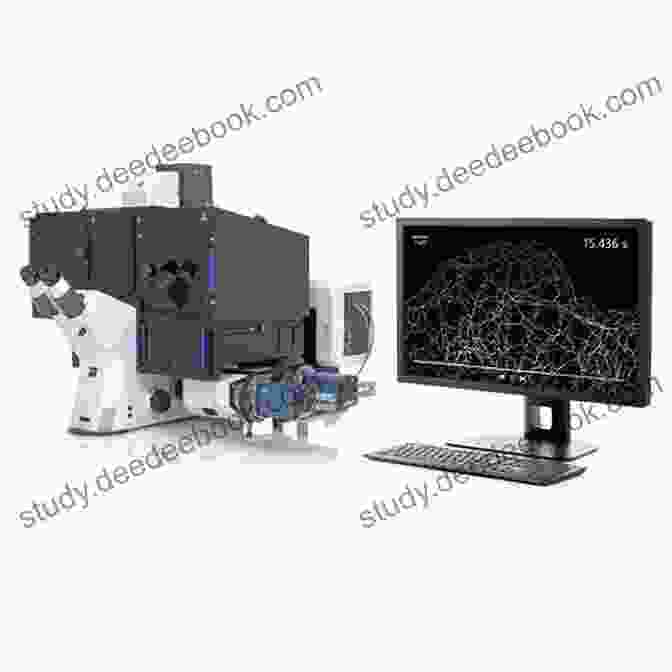
Super-resolution microscopy
Super-resolution microscopy techniques, such as stimulated emission depletion (STED) and photoactivated localization microscopy (PALM),overcome the diffraction limit of conventional microscopy. These techniques achieve sub-diffraction resolution, allowing for the visualization of fine cellular structures and interactions at the nanoscale.
Applications of Microanatomy
Microanatomy and its associated techniques have diverse applications in various fields:
1. Basic and Clinical Research
Microanatomy serves as a foundation for understanding the normal structure and function of organs and systems. It also plays a crucial role in investigating disease mechanisms and developing new therapies.
2. Pathology
Microanatomy is essential for diagnosing and classifying diseases based on tissue biopsies. Histological examination can reveal abnormal cellular changes, tissue damage, and the presence of microorganisms.
3. Forensic Science
Microanatomy is used in forensic investigations to examine evidence, such as hair, fibers, and blood, to identify and compare individuals or determine the cause of death.
Microanatomy, with its diverse approaches and techniques, provides a comprehensive understanding of the microscopic structure of biological tissues. From basic histological techniques to advanced imaging technologies, these methods empower scientists and clinicians to explore the intricate world of cells and tissues, unraveling the mysteries of health and disease. Microanatomy continues to evolve, offering exciting avenues for future research and applications.
5 out of 5
| Language | : | English |
| File size | : | 161686 KB |
| Text-to-Speech | : | Enabled |
| Screen Reader | : | Supported |
| Enhanced typesetting | : | Enabled |
| Print length | : | 1573 pages |
Do you want to contribute by writing guest posts on this blog?
Please contact us and send us a resume of previous articles that you have written.
 Book
Book Novel
Novel Page
Page Chapter
 Reader
 Library
 Paperback
 E-book
 Magazine
 Paragraph
 Sentence
 Bookmark
 Glossary
 Bibliography
 Preface
 Footnote
 Manuscript
 Codex
Codex Bestseller
 Classics
 Library card
 Autobiography
 Memoir
 Encyclopedia
 Dictionary
 Thesaurus
 Resolution
 Librarian
 Card Catalog
 Borrowing
 Archives
 Lending
 Journals
 Interlibrary
 Study Group
 Thesis
 Dissertation
 Storytelling
 Theory
 Textbooks
 Daniel Fedele
 Mark Victor Hansen
 M J Rose
 Ross Bentley
 Tim Parsons
 K G Reuss
 Patricia Hubbell
 M Never
 Terrence J Sejnowski
 Jean S Mill
 Paul Oyelakin
 Paul Smith
 John Russell Brown
 Regena Thomashauer
 Mouni Sadhu
 Nancy Radke
 Melissa Forti
 Joseph O Connor
 Meghan Quinn
 Susan Apps Bodilly
Light bulbAdvertise smarter! Our strategic ad space ensures maximum exposure. Reserve your spot today!
 Percy Bysshe ShelleySlow Travels with Georgia Lyn Wilkerson: Reconnecting with the Present Moment
 Jean BlairNational Geographic Readers: Animals in the City L2: Wildlife in the Concrete...


 Sammy PowellFollow ·10.2k
 Isaiah PriceFollow ·4.5k
 Jonathan FranzenFollow ·11.1k
 Tennessee WilliamsFollow ·16.2k
 José MartíFollow ·11.1k
 Ivan TurgenevFollow ·16.8k
 Kurt VonnegutFollow ·6.1k
 Chase MorrisFollow ·7.9k

 Dominic Simmons
Icky Island: An Unforgettable Adventure for Kids!
Introducing Icky Island: A Delightful One...

 Edward Reed
 Carlos Fuentes
The Midnight Breed: Embracing the Shadows and Unlocking a...
Welcome to the captivating world of...
 Ike Bell
Twelve Steps Toward Political Revelation: A Path to...
Politics, often perceived as a complex and...

 Cameron Reed
Travels in Arizona Goldfield: Unraveling the Threads of...
Nestled amidst the rugged...
 John Grisham
Flashpoints of Cinema History and Queer Politics:...
The relationship between cinema history and...
5 out of 5
| Language | : | English |
| File size | : | 161686 KB |
| Text-to-Speech | : | Enabled |
| Screen Reader | : | Supported |
| Enhanced typesetting | : | Enabled |
| Print length | : | 1573 pages |
